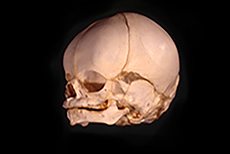
The Embryology of the Temporomandibular Joint
 This study examined 63 human fetuses, ranging in size from 4.5 to 255 mm crown-rump, to follow the embryological development of the temporomandibular joint. Sagittal, coronal, and transverse sections of the joint region were prepared and stained either in Milligan’s trichrome, a connective tissue stain, or in hematoxylin and eosin. The results of the study suggest that the lateral pterygoid muscle plays a pivotal role in the histodifferentiation of the temporomandibular joint. The lateral pterygoid muscle, with its fibrous insertions onto the condylar head, malleus, and temporal bone, directly contributes to the formation of the joint’s disk. The lateral pterygoid may also contribute indirectly, by mechanical stimulation, to the endochondral differentiation of the condyle and the formation of the inferior and superior joint cavities. The authors suggest that the early connection between the lateral pterygoid, condylar blastema, and Meckel’s cartilage which persists throughout the development of the temporomandibular joint may serve a function other than simply reflecting a phylogenic past when the malleus served as part of the mandibular joint.
To read more, click here.


